EnvisioningHealth - Changing Lives One Idea at a Time | InnovationXHealing
A robotic step forward in kidney preservation
NUH introduces a surgical technique using cheek tissue to restore kidney function,
circumventing the need for more invasive and complex procedures.
Issue 4 | March 2024
 Subscribe and ensure you don't miss the next issue!
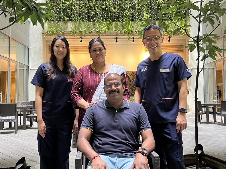
With the helping hand of a surgical robot, surgeons at the National University Hospital (NUH) have successfully completed a novel procedure designed to preserve a patient’s kidney function. The surgery involved the use of tissue from the patient’s mouth to mend a damaged ureter, the duct responsible for transporting urine from the kidney to the bladder.
The surgery was performed in September 2022 and the team only announced this after a cautious monitoring period of a year—a measure taken to ensure the stability of the graft.
Solutions in unexpected places
Led by
Associate Professor Tiong Ho Yee, Senior Consultant in the
Department of Urology, NUH, and
Dr Melissa Tay, Consultant in the same department, the surgery was carried out after the patient underwent two unsuccessful procedures, leading to a narrowing of his ureter in a condition known as ureteral stricture. This condition can result in infections, stone formation and kidney complications.
“In light of the increasing complexity posed by scar tissue build-up from previous surgeries, we turned to an innovative procedure known as buccal mucosa pyeloplasty,” says A/Prof Tiong. “First, we harvested the cheek’s lining, known as the buccal mucosa, as the graft material.”
“The buccal mucosa was selected as it closely resembles the lining of the urinary system, making it an ideal material for such repairs,” adds Dr Tay, explaining that a small piece of mucosa is highly stretchable and retains moisture well.
Following this, the surgeons cut open the constricted section of the ureter and sutured it with the harvested tissue via robotic laparoscopic keyhole surgery. To ensure adequate blood supply to the graft, the omentum, a membranous layer of fatty tissue that supports organs in the lower abdomen, was stitched onto the repaired section.
Technology lends a helping hand
The integration of a surgical robot was pivotal in this operation.
“Employing minimally invasive robotic techniques enabled us to achieve enhanced precision, reduce the risk of infection and facilitate quicker recovery compared to traditional surgical methods,” explains A/Prof Tiong. “In this case, the patient was spared from undergoing a more complex and challenging surgery, which might have involved creating a new tube created from a segment of the intestine, usually requiring open surgery.”
The shift from conventional open repair methods to laparoscopic (operations performed via small incisions assisted by a camera) and robotic techniques marks a significant step forward in urological surgery. In particular, these techniques can be beneficial in addressing ureteral strictures arising from various causes, including prior surgeries, radiation exposure and endometriosis—a condition where tissue similar to the uterus lining grows externally.
“The integration of advanced robotic technology with traditional surgical methods is something that I look forward to, as it is already laying the groundwork for safer, more efficient surgical solutions, ” adds Dr Tay.
Like this article? Simply subscribe to make sure you don't miss the next issue of EnvisioningHealth!
