Care at NUH

Guide to Intensive Care Unit (ICU) and High Dependency (HD) Unit 

2025/10/30

The Intensive Care Unit (ICU) and High Dependency (HD) Unit are wards where critically ill patients are admitted and cared for by a team of specialised healthcare professionals. The team comprises doctors, nurses, respiratory therapists, physiotherapists, pharmacists, radiographers, social workers and support care staffs. 

General guide

Communications
A family spokesperson should be nominated to be the point of contact between the ICU/HD team and family members. The spokesperson will be updated about the condition of the patient by the ICU/HD team on a regular basis.

Consent for treatment 
Most basic procedures and treatment are planned with the overall care and management of the patient in mind, hence presumed consent is exercised.

However, the spokesperson will be required to give consent for certain procedures if the patient is sedated or unconscious (as with most ICU/HD patients). In such instances, our medical staff will explain the risks and benefits of the treatment and offer alternatives, if any. 

Medical equipment
The patient may be connected to multiple equipment so that the ICU/HD team can monitor their condition. Some of the common types of equipment include:

Physiologic monitor monitors the patient’s heart rate, heart rhythm, blood pressure and oxygen saturation.
Intravenous (IV) or invasive lines provide fluid, medications and nutrition to the patient and monitor blood pressure.
Pumps are used to ensure continuous delivery of medicine via the IV or invasive lines for patient who is on several types of medications. 
Ventilator helps the patient to breathe through their nose or mouth via a breathing tube. They will not be able to speak while on the ventilator but this is only temporary.
Oxygen therapy is provided for patient through a face mask or nasal cannula.
Drains help to remove the patient’s excess body fluids. 

Most of the monitors and equipment are equipped with alarms to remind the staff of routine changes. As such, there is generally no cause for concern if the alarm sounds.

Visiting guidelines

Family members play an important role in the support and healing process of the patient. However, adequate rest and treatments are also important for a speedy recovery. Please observe the following visiting guidelines so that our patients can recuperate in a conducive environment.

The visitor policy is subject to changes in accordance to prevailing Ministry of Health (MOH) guidelines.

Visiting hours: 12pm – 8pm daily

Number of visitors: 
Please limit the visit to 2 visitors at any time to allow adequate rest and treatment of your loved one. 
Children who are 12 years and below will be denied entry.

Privacy
Blinds or curtains are drawn to protect the privacy of the patients. Please seek healthcare personnel permission before entering.

Medical care
Family member/caregiver may be asked to proceed to the visitors’ lounge during the doctors’ or nurses’ rounds and procedures.

Infection prevention
Proper hand hygiene is the best way to prevent and spread of infection. Family member/caregiver should use the antiseptic solution provided BEFORE and AFTER visiting the patient. Family member/caregiver who is feeling unwell (e.g. fever, cough or cold) should refrain from entering the ICU/HD.

Family/caregiver support services
We recognise the important role that family and caregivers play in supporting the care team. We also know that family member/caregiver, too, will need support, reassurance, and information.

Family member/caregiver may approach our staff if they require:  

  • Counselling
    For professional counselling and support by our Medical Social Workers
  • Financial counselling
    For financial assistance or information on patient’s hospitalisation bill
  • Spiritual needs
    We encourage you to approach our staff if you need support for your well-being, mental health, or social concerns. Our professional team is available to provide a confidential and supportive environment for discussion. Please don't hesitate to reach out if you would like to speak with someone.
Pain management

Pain after surgery is commonly experienced at the operative site. The pain can cause discomfort and delay the recovery process, prolonging the hospital stay.

Good pain management is important to relieve or reduce pain to provide greater comfort and allow the patient to engage in the following activities which help in faster recovery and earlier discharge:

  • Perform deep breathing exercises that can prevent the development of chest infection and other problems
  • Sit out of bed and walk

What can the patient do to manage pain?
Before surgery, the patient should try to understand the nature of his/her operation. Being well informed about the type of operation will help reduce anxiety.

Here are some useful questions the patient can ask the doctor or nurse before the surgery:

  • Will there be a lot of pain after surgery?
  • How long is the pain going to last?
  • What are the types of analgesic (pain-relief medication) which will be given after surgery?
  • What are the side effects of the pain-relief medication?
  • How often will the pain-relief medication be given?

Tips on how the patient can manage the pain after the surgery:

  • The patient should ask for the pain-relief medication as soon as he/she feels slight discomfort as it takes time for the medication to take effect.
  • The patient should take the pain-relief medication before the start of any activities such as physiotherapy as it is harder to ease the pain once it has set in.
  • The patient can help the doctor or nurse better manage their pain by providing an indication of how much pain they are experiencing using a pain scale. This allows the doctor and nurse to know the patient’s level of comfort and assess if the amount of pain relief medication administered is sufficient and adjust the treatment plan accordingly.

Pain-relief medications 
There are different types of pain-relief medications with different methods of administration to control pain after surgery.

Types of pain-relief medications

  1. Non-steroidal anti-inflammatory Drugs (NSAIDS) reduce swelling and soreness and are used to relieve mild to moderate pain.
  2. Opioids relieve moderate to severe pain.
  3. Local anaesthetics relieve severe pain. They are given either near the operative site or through a small tube in your back.

Methods of administration

  1. Oral (tablet/capsule)
    Common oral medicines are paracetamol, naproxen and tramadol)
  2. Injections
    a) Muscles
    The pain-relief medication is injected into the muscle of the upper arm, thigh or buttock. The common medicine used is pethidine, for relief of moderate to severe pain.
    b) Vein
    The pain-relief medication is given through a small tube inserted into the vein. Morphine is commonly used, and it is to treat moderate to severe pain. The pain-relief medication can also be administered continuously with the use of a syringe pump or a patient-controlled analgesia (PCA) pump. If the PCA pump is used, the patient will be able to self-administer the pain-relief medication whenever he needs it. 
    c) Back (epidural)
    Medicine is given through a small tube inserted into the patient’s back after numbing the skin with a local anaesthetic. The tip of the tube is inserted just outside the sack containing the spinal fluid and nerves. Local anaesthetics and opioids can be given through the tube for up to three days after surgery to provide a band of numbness over the operative site.
  3. Nerve block
    Pain-relief medication is given through a small tube inserted near the operative site. The common medicines used are local anaesthetics. It can be given up to three days after surgery to provide numbness over the operative site. 

Tips on how patients can manage mild to moderate pain:

  1. Support the wound site
    Coughing, sneezing or other simple essential movements like sitting up or getting out of bed can be painful. Supporting the wound site during these activities will help to reduce the pain.
  2. Apply cold pack
    Place well-wrapped cold packs on the area where pain is felt when ordered by doctor. Keep it for about 15 to 20 minutes so that it does not irritate your skin. It is safe to use and has no side effects.
  3. Acute Pain Service
    Our Department of Anaesthesia provides Acute Pain Service to help patients with moderate to severe acute post-operative pain. When you are admitted for surgery, our specialists will discuss the most effective strategies to manage your postoperative pain. We strive to address pain management comprehensively as part of your overall care plan. If pain persists despite initial treatments, our dedicated Pain Service team may review your case to optimize pain management and ensure your comfort during recovery.  
Brain aneurysm

A brain aneurysm is a swollen bulge or “ballooning” in the wall of a blood vessel in the brain. An aneurysm is a very serious, life-threatening emergency that needs to be treated right away. It leads to a bleeding (haemorrhagic) stroke. A “stroke” is a term doctors use when a part of the brain is damaged because of a problem with the blood vessels in the brain. 

Signs and symptoms of a brain aneurysm
Most brain aneurysms have no symptoms. But a large aneurysm can cause symptoms that include:

  • Severe headache
  • Pain in the face
  • Blurry or double vision

If the aneurysm ruptures, symptoms can include:

  • Sudden, severe headache - people may describe it as the worst headache they ever had
  • Stiff neck
  • Nausea and vomiting
  • Double vision
  • Drowsy
  • Coma  

Factors which may increase one’s risk of having a brain aneurysm

  • Having strong family history
  • High blood pressure
  • Smoking
  • Drinking too much alcohol
  • Using illegal drugs, such as cocaine or amphetamines
  • Taking diet pills

Tests to diagnose a brain aneurysm
The doctor may perform several different tests to diagnose brain aneurysm for patients who are displaying the symptoms:

  • Imaging tests
    CT scan and an MRI. Both show pictures of your brain. During these tests, you may be given an injection of a dye, which makes it easier for doctors to see the blood flow in the brain.
  • Cerebral angiogram
    For this test, your doctor will insert a thin plastic tube into a large blood vessel at the groin. Then, the tube will be advanced through your blood vessels past your heart to your brain. Your doctor will then inject a dye which will show up on the x-ray. This will allow the doctor to see the blood vessels in your brain to find the aneurysm.

Treatments for brain aneurysm 
The treatment of brain aneurysm is very critical, and the best option of treatment is entirely based on the patient's underlying condition, type and size of aneurysm, as well as a collaborative decision-making process between the neurosurgeon, interventional radiologist and patient.

  • Surgical clipping
    In this surgery, your doctor will place a tiny metal clip on the base of the aneurysm to stop the blood flow to it.
  • Endovascular coiling
    The first part of this treatment is the same procedure as a cerebral angiogram (see above). On top of injecting dye into the tube, your doctor will insert a special soft wire through your blood vessels into the aneurysm sac. The wire will then detach tiny titanium coils inside the aneurysm sac and seal it off from the blood vessel. 
How should the patient prepare for the surgery?
Before surgery, preparations that need to be done include:
  • Pre-operative investigations such as chest x-ray, electrocardiogram (ECG) and blood tests.
  • No food and drinks for at least 12 hours.
  • Decisions regarding treatment are made by the patient when capable. In cases where the patient is incapacitated, two consultants will collaboratively assess and determine the appropriate course of action. It's important to note that no written consent is required from next of kin in these situations.

What can the patient expect immediately after surgery?

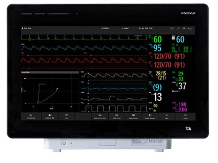
  • The patient may be required to stay in ICU or HD facility for close monitoring.
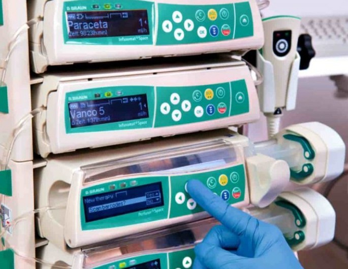
  • Presence of wound dressing, intravenous drip and head drain.
  • Presence of urinary catheter
  • Hourly monitoring of urine output
  • Frequent monitoring of vital signs and head chart.
  • For diet intake, there will be a gradual increase from nothing by mouth to diet of your choice.
  • Patient will be given painkillers for pain relief.
  • In surgical clipping, patient’s hair will be shaved off for the doctor to access the area to be operated on.
Length of hospitalisation
The length of hospitalisation for patients who have suffered subarachnoid haemorrhage is dependent on patient’s condition upon admission, and the treatment of concomitant issues such as cerebral vasospasm, hydrocephalus and respiratory issues. In patients with severe subarachnoid haemorrhage, hospitalisation may be up to three to four weeks.

Important points to note after surgery
  • Patient should not start doing any strenuous activities such as running, climbing, swimming until they have received clearance from their doctor to do so.
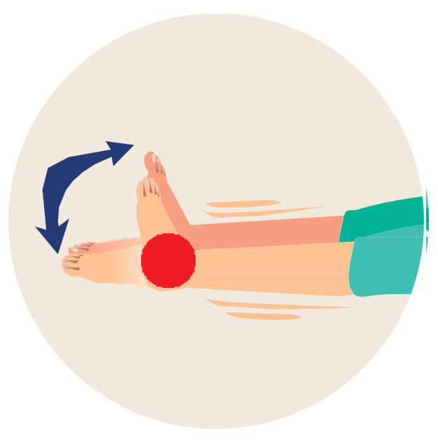
  • Patient should do light exercises as taught by the physiotherapist.
  • If patient is discharged with a dressing on the surgical site, they should get an outpatient appointment for removal of stitches before leaving the hospital.
  • Patient should not wet the dressing during shower. If the dressing gets wet, patient can go to the nearest clinic/polyclinic to have the wound cleaned and dressed.

If the patient experiences any of the following symptoms, proceed to the NUH Emergency Department immediately:

  • Signs of infection such as fever equal to or more than 38.0 degrees
  • Pain at the surgical site
  • Swelling, redness or leaking from the surgical site
  • Headache, vomiting, weakness, seizures, blurred vision and drowsiness.
Fall prevention

It is important that patients work together with the care team to prevent falls and possible injuries, which may prolong their hospital stays. If the patient falls, they may experience bleeding in the brain or other organs, broken bones or even go into a comma. The family/caregiver of the patient also plays a vital role in ensuring their safety. 

Tips for the family member/caregiver:

  • If patient is confused, stay with him/her at all times is strongly encouraged
  • Press the call bell and wait for assistance 
  • Frequently used items should be placed within reach of the patient.
  • Ensure bed rails are raised on both sides of the bed.
  • Take the time to perform lower limb exercises with the patient while in bed
  • The patient should not get out of bed alone if he/she feels weak or dizzy.

Patient should perform these limb exercises as instructed to prevent loss of lower limb strength:

Exercise 1: Quad sets

  1. Straighten your leg.
  2. Tighten* your thigh muscle and count to 10.
    *The back of knee should press against the bed if done correctly.
  3. Perform 5 sets.
    (Rest for 10 seconds between each set)
  4. Repeat twice a day

Exercise 2: Ankle pumps

  1. Move your ankle up and down 10 times.
  2. Perform 5 sets of 10 repetitions.
  3. Repeat twice a day.

Tips for the patient while walking:

  • Request for assistance from the nurse
  • Wear glasses and/or hearing aid if required
  • Use the recommended walking aid if required
  • Ensure that the pants are of suitable length and fold up the ends if they are too long
  • Wear appropriate non-slip footwear

Ways to the manage risk factors of falls

  1. Confusion
    Family member/caregiver should:
    - Accompany patient at all times, if possible
    - Keep bed rails up at all times
    Note: Hospital staff may apply restrainers to patient if necessary

  2. Drowsiness from certain medication or effects of anaesthesia
    Patient should:
    - Stay in bed with bed rails up at all times
    - Not climb out of bed
    - Press the call bell and wait for assistance

  3. Giddiness due to low or high blood pressure
    Patient should:
    - Change to sitting or standing position slowly
    - Sit down immediately if feeling giddy
    - Press the call bell and wait for assistance

  4. Urgent/frequent need to use the toilet
    Patient should:
    - Discuss toileting needs with nurse (e.g. voiding time, use of commode/urinal)
    - Press the call bell and wait for assistance if he/she experiences any weakness

  5. Difficulty in standing/walking
    Patient should:
    - Stay in bed with bed rails up at all times
    - Use walking aid as instructed
    - Not lean on movable items
    - Press the call bell and wait for assistance
High Flow Nasal Cannula (HFNC)

High Flow Nasal Cannula (HFNC) is an oxygen therapy that provides continuous heated and humidified oxygen through a nasal cannula placed in the nostrils. It helps to improve breathing by creating a small amount of positive pressure in the upper airway and providing a highly concentrated oxygen therapy. 

It helps the patient to:

  • Clear the secretion easily due to humidified air
  • Communicate while on HFNC
  • Eat and drink while on HFNC

HFNC is an alternative therapy for the patient who prefers non-invasive ventilation.

What kind of patient is suitable to undergo HFNC therapy?
  • Alert and able to protect their own airway
  • Free from facial injuries

What should the patient look out for during the HFNC therapy?
The nurse may apply a foam dressing over the patient’s cheeks and ear bridge to prevent any pressure injury. The patient may feel discomfort due to the strong air flow that goes into their nostrils so they should give their body some time to adapt. 

Things patient to take note of while undergoing HFNC therapy
Do’s

  • Comply with treatment.
  • Breathe through the nose.
  • Patient may remove nasal cannula temporarily to clear mucus.
  • Patient may adjust nasal cannula to one’s comfort while ensuring it is in the nostrils.
Don’ts
  • Patient should not remove the nasal cannula by themselves for prolonged period or without the doctor’s advice.
  • Patient should not raise the tubing of the HFNC as the fluid accumulated in the tubing may enter the nose and lungs.
Ventriculo-Peritoneal Shunt (VP Shunt)

Ventriculo-Peritoneal shunt (VP shunt) is used to drain out excess fluids circulating around the brain and spinal cord canal. This fluid is also known as cerebrospinal fluid (CSF). If there is too much of this fluid in the ventricles of the brain, it will increase the pressure within the brain. This condition is known as hydrocephalus.

Signs and symptoms of hydrocephalus:

  • Headache
  • Drowsiness
  • Nausea and vomiting
  • Memory loss
  • Confusion
  • Difficulty in balancing and walking
  • Loss of ability to control urination

How does a VP shunt work?
A VP shunt is a long, plastic tube that drains excess CSF from the brain to another part of the body. This drainage prevents the increase in pressure on the brain caused by hydrocephalus. The symptoms should be relieved immediately or over a few weeks. 

There are at least three parts to a VP shunt:

  • The first part is the ventricular catheter, which goes into the brain.
  • The second part is the valve, which controls the pressure within the brain.
  • The third part is the distal catheter, which is underneath the skin and connects the other part(s) of the VP shunt to a space within the body, usually the abdominal cavity (also called the peritoneal cavity). Fluid will drain from ventricle to abdomen and will be absorbed into your general circulation.

The amount of fluid that is drained depends on the valve pressure. Most valves are set to a specific setting and cannot be changed. However, there are valves that can be programmed to a variety of pressures which is called Programmable Shunt. Your neurosurgeon will select the type of valve shunt that is most suitable for you.

Warning signs that a shunt is not working properly
VP shunt can last a lifetime. However, shunt obstruction due to blockage can happen. 

Signs of a VP shunt malfunction:

  • Headache
  • Vomiting
  • Blurred vision
  • Seizures
  • Decrease in conscious level

If you have any of the above symptoms, please consult your doctor or visit the NUH emergency department immediately.

VP shunt infection
A shunt infection occurs when bacteria infect the tissues surrounding the VP shunt which can lead to malfunction of the shunt. A shunt that does not work well can cause an increase in pressure within the brain. This increase in pressure can damage the brain.

Signs and symptoms of a VP shunt infection:

  • Persistent high fever (equal to or more than 38.0°C)
  • Pain around the shunt or around the shunt tubing
  • Irritability or personality changes
  • Swelling or tenderness of the abdomen with a VP shunt
  • Persistent nausea and vomiting
  • Headaches
If you experience any of the above signs and symptoms, please visit the NUH emergency department as soon as possible.
External Ventricular Drainage (EVD)

External Ventricular Drainage (EVD) is a temporary drainage of cerebrospinal fluid (CSF) from the fluid-filled cavities of the ventricles in the brain or the lumbar space of the spine into an external collection bag.

EVD is needed when there is an increase of fluid in the brain caused by an obstruction, infection or injury involving the brain. This increases pressure in the brain as the normal flow of fluids is compromised. To reduce the pressure, placement of an external drain is done.

EVD is a temporary way to drain CSF from the ventricles to relieve pressure in the brain.

Things patient should take note of while undergoing EVD:
Do’s:

  • Inform the nurse if the patient wants to change his/her sitting position.

Don’ts:

  • Don't touch the EVD drain without supervision.
  • Don't touch EVD wound or the surgical site.
  • Don't adjust the EVD chamber.
  • Don't move the bed without the presence of a nurse.
  • Don't get out of bed without informing the nurse.  
Awake Prone Positioning

Awake prone positioning is a therapeutic therapy where the patient will be lying on their stomach. It aims to improve their oxygen level and ease breathing. 

How does awake prone positioning help the patient?

  • Lying on the stomach and in various positions helps air enter all areas of the lungs.
  • Patient may notice improvement in breathing immediately or several minutes after each position change.
  • It is a safe and rapid implementation to prevent respiratory failure from progressing.
  • Literature has shown that it can contribute to low mortality and high discharge rates among patients if started early.

What kind of patient is suitable to do awake prone positioning?

  • Fully alert and be able to communicate and cooperate with procedure
  • Able to rotate and adjust position independently
  • No unstable spine/chest/abdominal/facial surgeries or injuries
  • Not morbidly obese 
  • Not in 2nd/3rd trimester of pregnancy

What do patient need to prepare for awake prone positioning?

  • Pillows: Place under the chest, hips and legs where required for comfort
  • Empty the bladder and bowel and ensure personal hygiene
  • Place necessary belongings (e.g., handphone, tissue box) within reach 

Patient should cycle through the following positions and repeat the cycle as often as per the doctor’s advice (at least 2 to 4 times a day)


(Credit: Krames, 2020)

Things patient to take note of while doing awake prone positioning
Do’s:

  • Rest for one hour after a meal before proning, to prevent regurgitation
  • Maintain each position for at least 30 minutes and up to 2 hours
  • Adjust the position regularly and gently to ensure comfort and prevent pressure injury
  • Deep breathing exercise and use incentive spirometry
  • Inform the nurse if the patient experiences increased difficulty in breathing, or discomfort/pain over any pressure points

Don’ts

  • Patient should not spend too much time lying flat on their back
  • Patient should not stay in any position that causes discomfort or pain

ICU Delirium

ICU delirium is a common and serious complication that affects many critically ill patients in intensive care units. It is characterized by acute changes in mental status, including inattention, disorganised thinking, and altered levels of consciousness. Delirium can lead to increased mortality, longer hospital stays, and long-term cognitive impairment.

Last updated on
Best viewed with Chrome 79.0, Edge 112.0, Firefox 61.0, Safari 11
National University Health System
  • National University Hospital
  • Ng Teng Fong General Hospital
  • Alexandra Hospital
  • Tengah General and Community Hospital
  • Jurong Community Hospital
  • National University Polyclinics
  • Jurong Medical Centre
  • National University Cancer Institute, Singapore
  • National University Heart Centre, Singapore
  • National University Centre for Oral Health, Singapore
  • NUHS Diagnostics
  • NUHS Pharmacy
  • NUHS Regional Health System Office
  • NUS Yong Loo Lin School of Medicine
  • NUS Faculty of Dentistry
  • NUS Saw Swee Hock School of Public Health
Back to Top