NUH will NEVER ask you to transfer money or disclose bank details over a call. We also do not endorse, sell or promote products on social media. If in doubt, call the 24/7 ScamShield helpline at 1799, or visit the ScamShield website at www.scamshield.gov.sg. For more information, please refer to: https://www.nuh.com.sg/beware-of-scams-impersonating-nuh
The oesophagus or gullet (食道 in Mandarin) is a muscular tube that transmits food from the mouth into the stomach. Once food enters the oesophagus, it is transmitted down to the stomach through a series of muscular contractions — a process called 'peristalsis'.
When food passes from the lower end of the oesophagus into the stomach, it encounters a special muscular ring known as the lower oesophageal sphincter (LES). When the LES relaxes, the food will enter the stomach. The LES is important because it prevents the backflow of food and acid from the stomach up into the oesophagus.
On the other hand, if the LES fails to relax, the food in the oesophagus is unable to enter the stomach. Over time, the motor function of the oesophageal muscle weakens and the oesophagus enlarges. This failure of relaxation of the LES occurs in a disease called achalasia.
Achalasia is a relatively rare disease affecting about 1 in 100,000 people. It affects both adults and children. Men and women are equally affected. The exact cause is unknown.
Achalasia is a chronic disease; most people suffering from this condition will try to tolerate the difficulty in swallowing and adjust their diet to take small portions or choose to have a liquid diet. Some of them may become thin and malnourished with poor quality of life. Nevertheless, this condition is treatable. Speak to your doctors or contact us through this website, email or telephone for further details.
Signs & symptoms
Difficulty swallowing
Food regurgitation
Night cough
Aspiration
Recurrent lung infections
Chest pain
Weight loss
Diagnostic and treatment options
Test and diagnosis
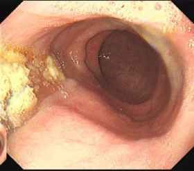
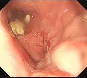
Upper endoscopy is a common test used to examine the upper digestive tract. The procedure is usually performed in an outpatient setting. It involves insertion of an endoscope, which is a thin flexible tube with a camera at the tip, to allow for visual examination of the oesophagus and stomach. The procedure takes less than 10 minutes and is often performed under light sedation. It is very useful for excluding any tumour or blockage of the oesophagus. In achalasia, the esophagus will be wider than usual, and is sometimes filled with residual food (Fig. 1a and Fig. 1b).
Fig. 1a
Fig. 1b
Another method of detection is using barium, a common X-ray contrast liquid. When swallowed, barium will outline the inner surface of the oesophagus, allowing the oesophagus to be seen on X-ray. The typical features of achalasia include a dilated oesophagus and narrowing at the end of the oesophagus, producing a 'bird beak' appearance.
Oesophageal manometry is the most important test to confirm the diagnosis of achalasia and allows us to see what happens when a patient swallows. A small thin tube with pressure sensors is inserted gently into the oesophagus and stomach. The movement of the oesophagus and pressure of the LES will be recorded to determine any impairment in function.
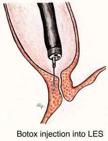
Botulinum Toxin Injection
Botulinum Toxin (commonly known as 'botox') blocks the release of chemicals in the muscle that induce contractions, hence resulting in relaxation of the LES. It is a safe and effective treatment for achalasia. Side-effects are minimal but relief is temporary.
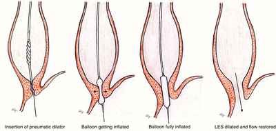
Balloon Dilatation
A balloon attached at the tip of a catheter is inserted into the lower oesophagus. The balloon is inflated, enlarging the passage of the lower oesophagus into the stomach. The LES's pressure will be reduced as the force of the expanding balloon tears its muscle fibres.
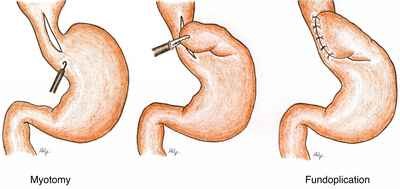
Laparoscopic Myotomy
Laparoscopic myotomy involves surgical division of the muscle of the LES. This procedure is done under general anaesthesia. It is a minimally invasive surgery. The muscle fibres of the LES are cut longitudinally from lower oesophagus to upper gastric (cardia).
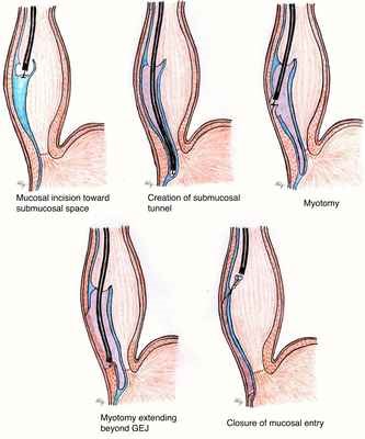
Peroral Endoscopic Myotomy (POEM)
This is the treatment for achalasia with no surgical incision required. The procedure is done endoscopically under general anaesthesia. The endoscope is inserted into the oesophageal lumen. A tunnel is created below the inner lining of the oesophagus all the way to the LES. The muscle fibres of the lower oesophagus and gastric cardia are then divided endoscopically.
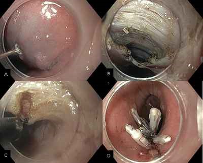
Endoscopic view of POEM.
A: 2cm longitudinal incision is made.
B: Submucosal tunnel is created leaving the underlying muscularis propria.
C: Circular muscle is cut, preserving the outer longitudinal muscle layer.
D: Clips are placed to close the initial mucosal layer.
A short video recording of the procedure can also be viewed here. Do note that this video contains images which some viewers may find disturbing.